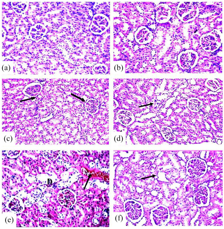
Figure 5.
Photomicrographs of sections in the kidney of rats in the different groups stained with hematoxylin and eosin (H&E), magnification ×400. (a) Control group. (b) PIC group showing normal histological structure of renal parenchyma characterized by circumscribe glomeruli with normal structure of capillary tufts and Bowman’s capsule. The renal tubules of both proximal and distal convoluted tubules showed intact epithelial lining with empty lumen (score 0). (c) RES group showing tubular epithelial lining with granulo-vacuolar degeneration (arrows) and narrowing of tubular lumen. The epithelial lining of both proximal and distal convoluted tubules lining showed necrosis and apoptosis (score 4). (d) RES + PIC group showing improvement of the kidney architecture with some swelling of tubular epithelial lining (score 1). (e) IR group showing leukocytic infiltration, degeneration in lying epithelium (D) of some tubules with congestion of blood vessels (score 4). (f) IR + PIC group showed mild swelling of tubular epithelial lining without significant pathological alteration (score 1).

