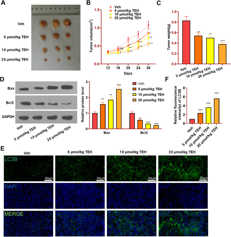
Figure 2.
TEH reduced HCC cells growth in vivo and induces apoptosis and autophagy. Human HepG2 cells were used to test the effect of TEH on HCC cell growth in vivo. HepG2 cells were subcutaneously injected in to the nude mice, which were then dealt with different doses of TEH (5-20 μmol/kg body weight). The tumor volume was measured every 4 days since the twelfth day of modeling. After a total of 28-day incubation, the mice were sacrificed and the tumors were isolated and weighted. (A) Tumor images. (B) Tumor volume. (C) Tumor weight. (D) Westrn blot was conducted to evaluate the apoptosis markers (including Bax and Bcl2) in the tumor tissues. (E-F) Tissue immunofluorescence was performed to detect LC3B (green marker) expression in the tumor tissues, and the fluorescence intensity of LC3B was analyzed. N = 5. **P < 0.01, ***P < 0.001 (vs. veh group).

