Figure 1.
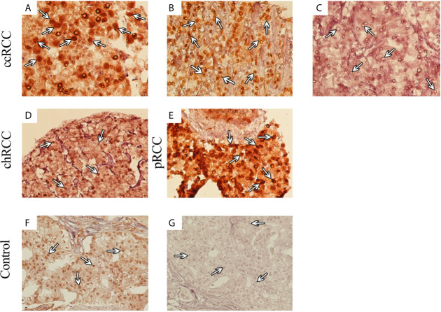
Immunohistochemical (IHC) Analysis of Nuclear SMAD4 Expression in Different 707 Renal Cell Carcinoma (RCC) Samples. RCC samples expressed SMAD4 at various levels. SMAD4 expression in clear cell RCC at various levels: weak (A), moderate (B), and strong (C). Moderate expression of SMAD4 in chromophobe RCC (D) and strong expression of SMAD4 in papillary RCC (E). IHC staining of breast cancer tissue as positive (F) and negative (G) controls. (A–C) are presented with a magnification of 400×, (D–G) are presented with a magnification of 200×).

