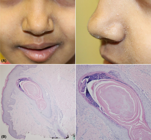
FIGURE 1.
Clinical and histologic images of trichodysplasia spinulosa. A, Multiple follicle-based papules on the central face with white dystrophic hairs protruding from the center of the papules. B, Biopsy lacked a well-defined hair shaft and showed a dilated infundibulum filled with keratinaceous and apoptotic debris

