Figure 5.

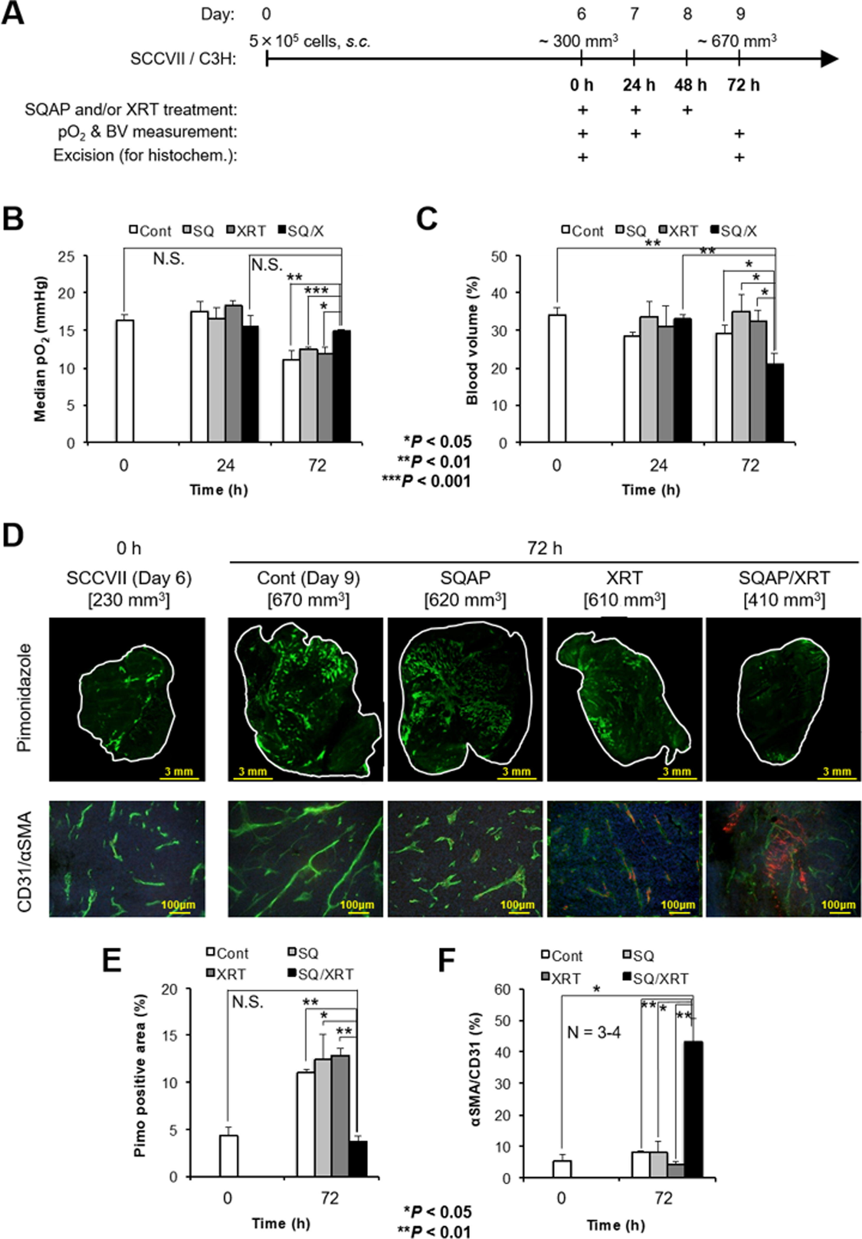
Monitoring of physiological properties in the tumors before and after 3 days of daily SQAP combination therapy with XRT. A, Schematic representation of the experiments. The 3 days (0, 24, and 48 h) of daily treatment with SQAP and/or XRT started at day 6, with a SCCVII tumor size of ~300 mm3. B, C, Noninvasive monitoring of tumor median pO2 (B) and blood volume (BV) changes (C) in SCCVII tumors before (0 h), and 24 or 72 h during or after the three treatments. B, The tumor median pO2 (mmHg) obtained from the EPRI pO2 map. C, BV (%) changes calculated using MRI with the blood pooling T2 contrast agent USPIO. Each pO2 and BV value was calculated from three 2 mm thick center images from each tumor (Supplementary data Figure S4). D–F, Immunohistochemical analysis of tumor slices before (0 h) or after (72 h) the 3 days of daily treatment. A dose of 60 mg/kg pimonidazole was intravenously injected as a hypoxic marker, and mice were excised 45–60 min after injection. Anti-CD31 antibody was used as a marker of vascular endothelial cells in the pathological tumor vasculature. Anti-α-SMA antibody was used as a marker for pericytes. E, Quantitative measurement of the pimonidazole-positive area (%) in whole center slices before (0 h) or after (72 h) three treatments. F, The ratio of α-SMA/CD31 expression (%) as a normalization index. Data are means ± SE (n = 3). *, P < 0.05, **, P < 0.01.
