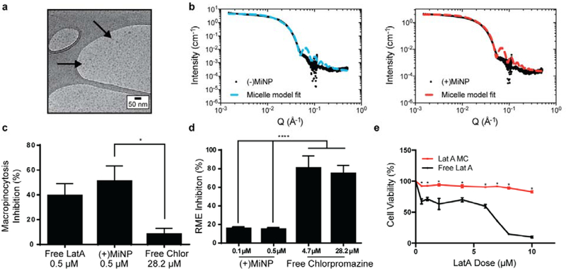
Figure 2: LatA retains its endocytic inhibition properties and does not change the size of PEG-b-PPS micelles when encapsulated.
a) Cryogenic transmission electron microscopy (CryoTEM) of MiNP visually confirms retention of micellar structures. b) MiNP with ((+)MiNP) and without ((−)MiNP) loaded LatA were characterized via small angle x-ray scattering (SAXS) and fitted with a micelle model fit using SASView. c) Free LatA and (+)MiNP significantly inhibited macropinocytosis by RAW264.7 macrophages as compared to clathrin-mediated endocytosis inhibitor chlorpromazine. Cells were treated with each inhibitor for 2 h followed by 30 min of incubation with pHrodo dextran prior to analysis by flow cytometry. Data are shown as a percentile scale of endocytosis inhibition. On this scale, 0% represents standard cell uptake with no inhibitor, while 100% represents complete inhibition with no uptake of dye. N=3 p<0.001. d) In comparison, uptake of transferrin conjugated pHrodo dextran by macrophages via receptor-mediated endocytosis (RME) was significantly inhibited by chlorpromazine compared to (+)MiNP. N=3 p<0.001. e) Loading within (+)MiNP significantly decreased the toxicity of LatA. Macrophages were incubated with various doses of free LatA or (+)MiNP for 4 h and assessed by flow cytometry for viability via the Zombie Aqua live/dead assay. N=3 p<0.05.

