Figure 2.
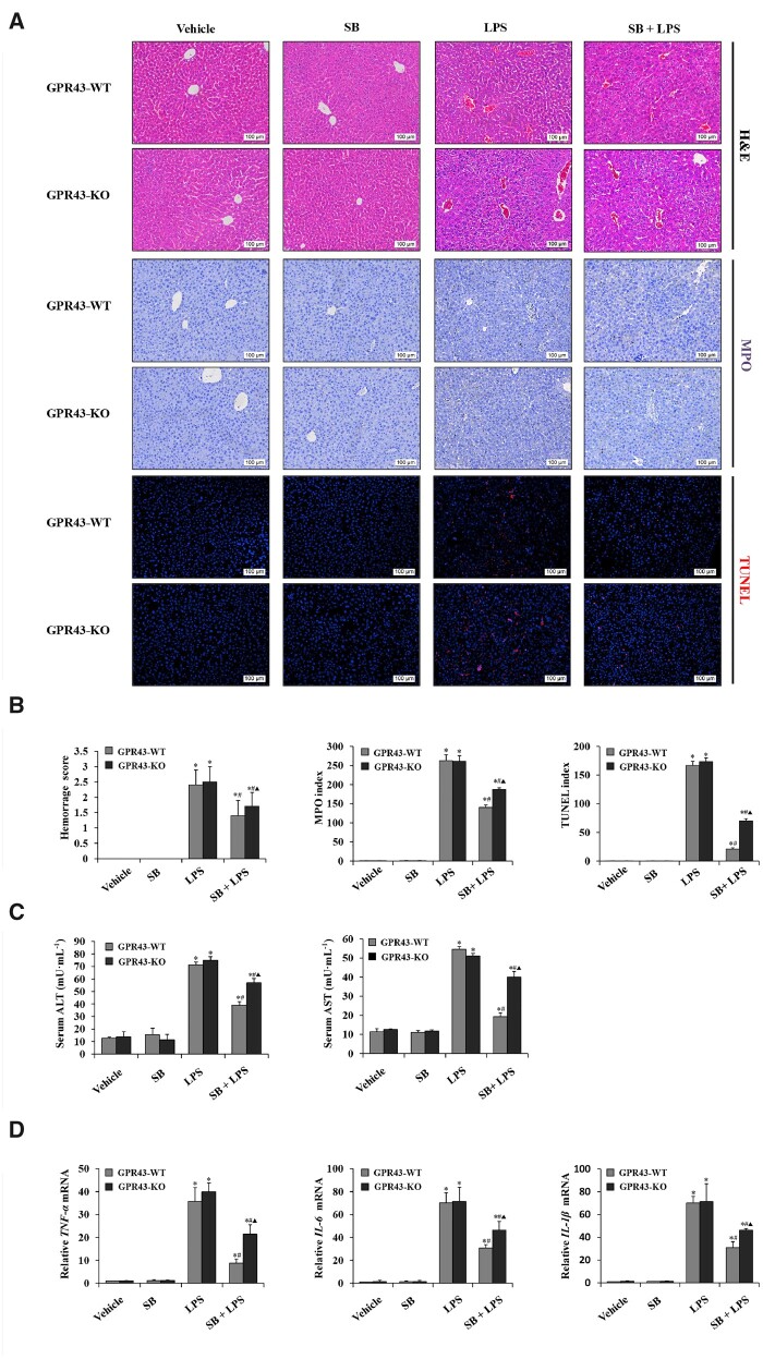
GPR43 deficiency interrupted the protection of sodium butyrate on LPS-induced liver injury and inflammation. (A) H&E staining, immunohistochemical staining for MPO, and TUNEL staining of liver tissue in female GPR43 KO mice and WT littermates treated with SB, SB + LPS, LPS, and normal saline (magnification, ×200). (B) Histology score of hemorrhage, MPO index, and the apoptosis index was calculated from TUNEL staining in female GPR43 KO mice and WT littermates treated with SB, SB + LPS, LPS, and normal saline. (C) Serum levels of ALT and AST were detected using ELISA kits. (D) RT-PCR was used to determine relative mRNA levels of TNF-α, IL-6, and IL-1β in liver tissues. *P < 0.05 vs vehicle group in the same type of mice; #P < 0.05 vs LPS group in the same type of mice; ▲ P < 0.05 vs SB + LPS group in GPR43 WT mice. Values are expressed as mean ± SD (n = 6 per group). MPO, myeloperoxidase; TUNEL, terminal deoxynucleotidyl transferase-mediated dUTP nick end labeling; SB, sodium butyrate; LPS, lipopolysaccharide; ALT, alanine aminotransferase; AST, aspartate transaminase; RT-PCR, real-time polymerase chain reaction.

