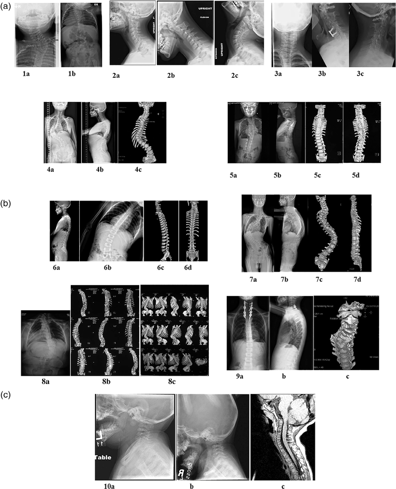
FIGURE 1.
Radiographic illustration of vertebral malformations seen in all probands. (a) Proband 1: Plain X-rays showing fused cervical and thoracic vertebrae in a 9-month-old male (1a, 1b). Proband 1 also have ASD. Proband 2, lateral neck films (2a) with flexion (2b) extension (2c) views showing fused cervical vertebrae in a 13-year-old male. Proband 2 also have macrocephaly, hydrocephalus, developmental delay, and intellectual impairment. Proband 3, 42-year-old male with dextrocardia, VSD. AP (3a), lateral (3b), and flexion (3c) X-rays showing fused cervical vertebrae. Proband 4, 12-year-old female with vertebrae deformation of C6, T3, T10, and segmentation defects of C3-C4, C6-C7, T3-T4 as shown in AP (4a) and lateral spine (4b) X-rays and AP 3D spine imaging (4c). Proband 5, AP (5a) and lateral spine (5b) X-ray and AP (5c) and PA (5d) 3D imaging of the spine showing extensive segmentation defects from C7-T9, thoracic spondylosis, tethered cord in 8-year-old male. (b) Proband 6, 10 year old male, AP (6a) and lateral (6b) X-ray and 3D spine imaging (6c and 6d) showing segmentation defects of C2/C3, non-segmented hemivertebrae of T4, and non-segmented wedged vertebrae of T12. Proband 7, 16-year-old male with segmentation defect of C6, non-segmented hemivertebrae of C7, and wedged vertebrae of T6/T7 as shown in AP (7a) and lateral (7b) X-ray and 3D imaging (7c and 7d). Leg-length discrepancy can be noted on the AP X-ray. Proband 8, 42-year-old female with C5-T5 segmentation defect with a short neck and non-segmented hemivertebrae of L1 as demonstrated by AP (8a) X-ray and 3D imaging (8b and 8c) of the spine. (i) Proband 9, spine C2-C7 segmentation defect in a 9-year-old male. AP (9a) and lateral spine X-ray (9b) and 3D imaging (9c) showing C-spine fusion. (c) Proband 10, lateral neck X-ray (10a and 10b) and MRI (10c) demonstrating incomplete segmentation of C1 fusion to Clivus, C3-C4, and hypoplastic occipital condyles

