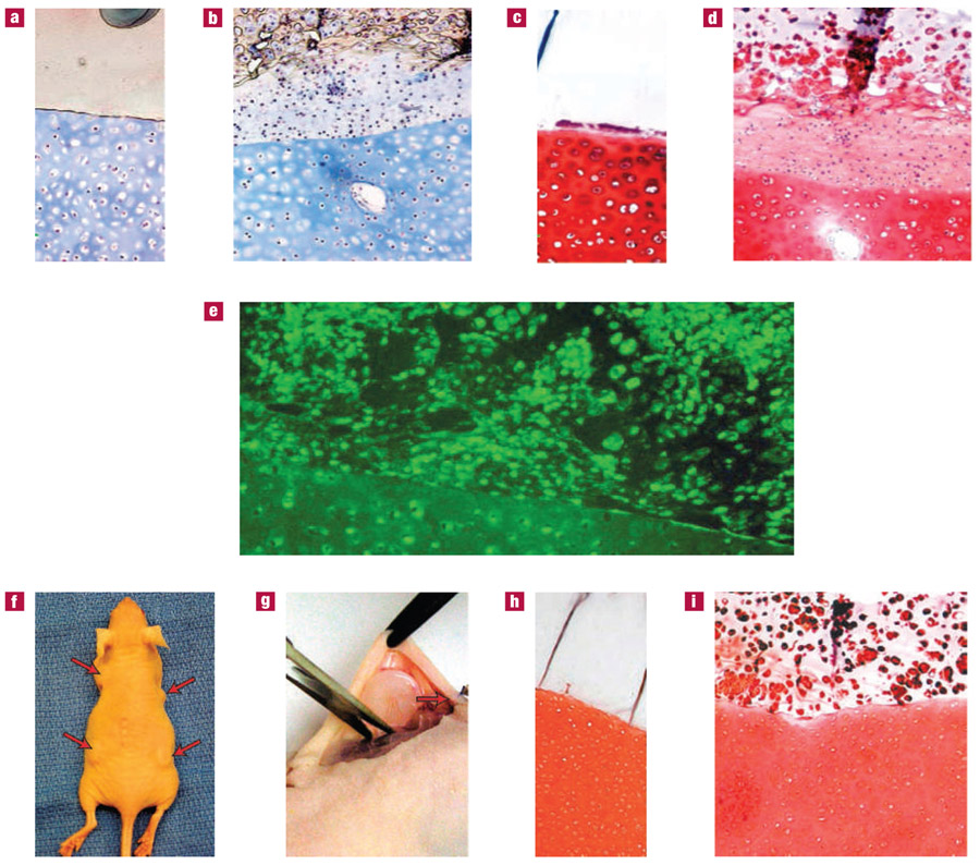
Figure 3. Integration of hydrogels and developing tissue to the cartilage surface with CS adhesive.
a–i, Hydrogel constructs were bonded to cartilage with the CS adhesive and incubated in vitro (a–e) or in vivo (f–i) for 5 weeks. Hydrogels without cells were integrated to cartilage explants and the resulting constructs were stained with Masson’s Trichrome (a) and Safranin-O (c) after in vitro culture to visualize collagen and proteoglycans, respectively. The hydrogels remained firmly attached to the cartilage tissue, which also retained cell viability and extracellular matrix. Hydrogels with encapsulated chondrocytes demonstrated total collagen (b) and proteoglycan (d) production typical of neocartilage. The neocartilage was firmly attached to the cartilage explant. Type-II collagen (e), specific to hyaline cartilage, was present in and around cells at the interface and within the native cartilage. Integrated cartilage–hydrogel constructs were also implanted subcutaneously in athymic mice (f) and collected after 5 weeks (g). Acellular hydrogels remained firmly attached to the cartilage surface without invasion of cells or extracellular matrix production (h). Similar to the case for in vitro incubation, the hydrogels containing cells produced neocartilage that was bound to the cartilage surface. Proteoglycans (I, Safranin-O) were present throughout the hydrogel layer and at the interface of the engineered and native cartilage tissue.

