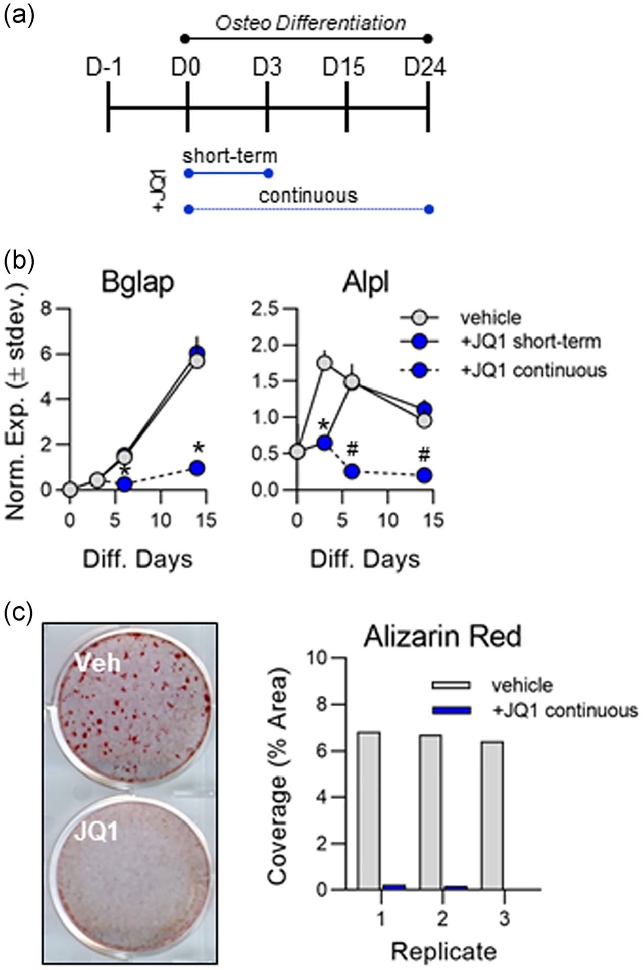
FIGURE 5.

Continuous +JQ1 treatment prevents osteogenesis of MC3T3 cells. (a) Experimental outline indicating short-term (3 days) and continuous 100 nm +JQ1 treatment in differentiating MC3T3 cells. (b) Quantitative reverse transcription polymerase chain reaction (RT-qPCR) analysis performed on messenger RNA (mRNA) collected throughout the osteogenic differentiation time course (n = 3, mean ± STD). (c) Representative image (left) and ImageJ quantification (right) of alizarin red staining for hydroxyapatite deposition on Day 24 of osteogenic differentiation. All error bars represent ± STD of three biological replicates. Statistical significance to vehicle (dimethyl sulfoxide [DMSO]) control group indicated as follows: *p < .01, #p < .001
