Fig. 6.
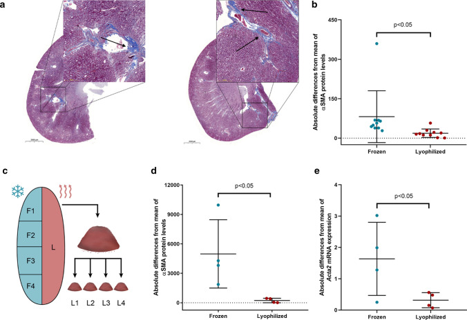
Comparison of scatter in frozen and lyophilized, pulverized samples from fibrotic kidneys. a Representative Masson's trichrome-stained sections of diabetic rat kidneys. Arrows show examples of focal fibrosis. (10–200 × magnification, scale bar = 2000 μm, 50 μm.) b Absolute differences of alpha smooth muscle actin protein (αSMA) values to mean in diabetic rat kidneys processed conventionally (Frozen) or homogenized after lyophilization (n = 10/group). c Schematic presentation of sample processing for homogeneity investigations. One half of each kidney was lyophilized (L) and pulverized, protein and RNA were isolated four times from homogenous powder (L1–L4). The other half went through conventional frozen sample processing; four pieces were cut for the isolations (F1–F4). d Absolute differences of αSMA protein and e alpha smooth muscle actin mRNA (Acta2) values to mean in frozen parts vs. lyophilized homogenous powder of the same diabetic rat kidney samples (n = 4 frozen parts and 4 powder portions of the same kidneys)

