FIGURE 1.
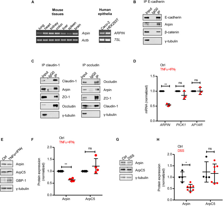
Arpin is downregulated under inflammatory conditions. (A) RT-PCR for Arpin and Actb using cDNA derived from the indicated mouse tissues (left) and ARPIN and 7SL as housekeeping gene using cDNA derived from the indicated human cell lines (right); n = 3. (B) Western blot of E-cadherin immunoprecipitates. β-catenin was probed as a positive control of interaction and γ-tubulin as a negative control; n = 3. Input = whole cell lysate; IgG0 = IP using IgG as control, IP = IP using specific antibodies. (C) Western blots of claudin-1 and occludin immunoprecipitates. ZO-1 was probed as a positive control and γ-tubulin as a negative control, n = 3. (D) Quantitative real-time RT-PCR for the Arp2/3 inhibitors ARPIN, PICK1, and AP1AR using cDNA from Caco-2 colon epithelial cells treated or not with tumor necrosis factor (TNF)α/interferon (IFN)γ (n = 3; two-tailed t-test). Data are shown as relative expression normalized to the housekeeping gene 7SL. (E) Western blot for arpin and the Arp2/3 subunit ArpC5 in Caco-2 cells treated or not with TNFα/IFNγ. Guanylate-binding protein-1 (GBP-1) was used as an inflammation positive control. (F) Densitometric analysis of panel (E) (n = 4; two-tailed t-test). (G) Western blot for arpin and ArpC5 in colons from control and dextran sulphate sodium (DSS)-treated mice. (H) Densitometric analysis of panel G (nCtrl = 4, nDSS = 7; two-tailed t-test followed by Mann–Whitney test). ns, non-significant; *p < 0.05; **p < 0.01.

