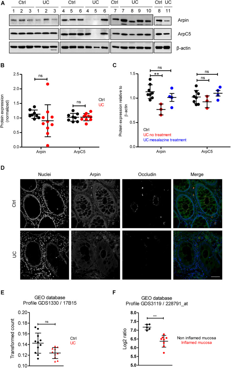
FIGURE 4.
Arpin is downregulated in inflamed areas of colon tissue from ulcerative colitis (UC) patients. (A) Western blot for arpin and ArpC5 from resection specimens of patients with UC and non-inflamed controls. β-actin was probed as the loading control. (B) Quantification of pixel intensities including all bands (compare Table 1 for single values; nCtrl = 8, nUC = 10; two-tailed t-test followed by Mann–Whitney test; ns, non-significant. We only excluded sample UC 5 as a technical outlier because of the absence of both arpin and ArpC5 and a much weaker β-actin band. (C) Comparison of pixel intensities from bands of control samples, samples from patients receiving no treatment, and patients receiving mesalazine in their medication regimen. **p < 0.01. (D) Representative immunostaining for occludin and arpin from resection specimens of patients with histologically active UC and non-inflamed controls; n = 3. Bar = 20 μm. (E) Arpin mRNA analysis in biopsies from UC patients compared to controls (n: Control = 11, UC = 10; one-way ANOVA with Kruskal–Wallis correction. (F) mRNA analysis of arpin from publicly available datasets (n: non-inflamed mucosa = 5, inflamed mucosa = 8; two-tailed t-test with Welch’s correction). ***p < 0.001. ns = not significant.

