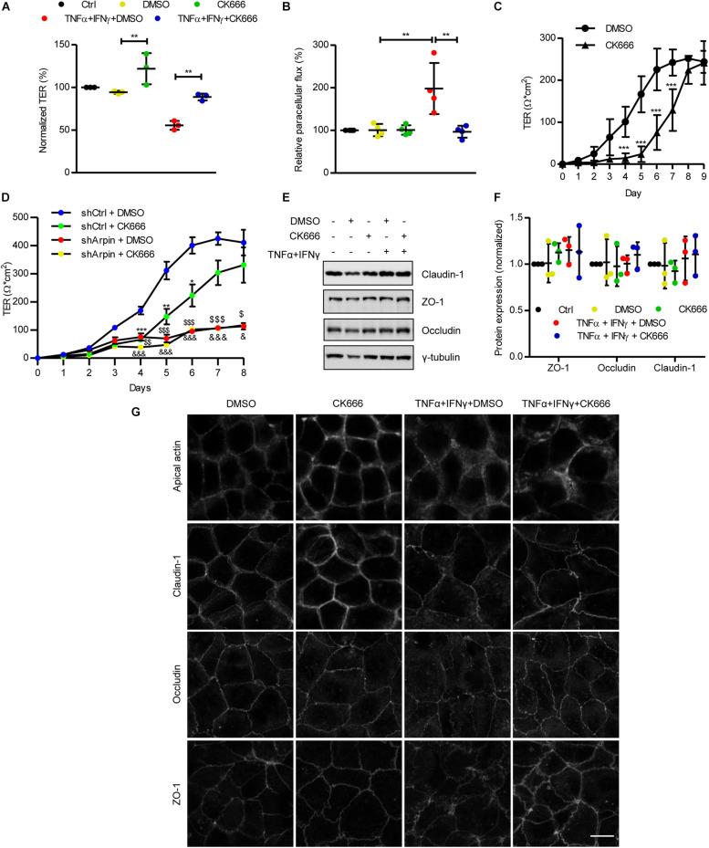
FIGURE 5.
CK666 reinforces the epithelial barrier. (A) Transepithelial electrical resistance (TER) measurements of confluent Caco-2 cells treated or not for 48 h with tumor necrosis factor (TNF)α/interferon (IFN)γ and CK666. The vehicle dimethylsulfoxide (DMSO) was used as control (n = 3; one-way ANOVA). (B) Paracellular flux of confluent Caco-2 cells treated or not for 48 h with TNFα/IFNγ and CK666 was measured 2 h after adding 4 kDa fluorescein isothiocyanate (FITC)-dextran; color code as in panel (A); n = 4; one-way ANOVA. (C) Time-course TER measurements of sparse Caco-2 cells in the presence or absence of CK666 (n = 5; two-way ANOVA). (D) Time-course TER measurements of sparse control and arpin-depleted Caco-2 cells in the presence or absence of CK666 (n = 6; two-way ANOVA). Data are compared with shCtrl DMSO-treated cells (*p vs. shCtrl + CK666, $p vs. shArpin + DMSO, and &p vs. shArpin + CK666). (E) Western blot for claudin-1, zonula occludens 1 (ZO-1), and occludin of Caco-2 monolayers treated or not with TNFα/IFNγ in the presence or absence of CK666. (F) Densitometry analysis of panel (D) (n = 3; one-way ANOVA with Bonferroni’s correction). (G) Confocal microscopy analysis of apical actin, claudin-1, occludin, and ZO-1 in Caco-2 cells treated or not with TNFα/IFNγ and CK666; n = 3. Bar = 10 μm. **p < 0.01; ***p < 0.001.

