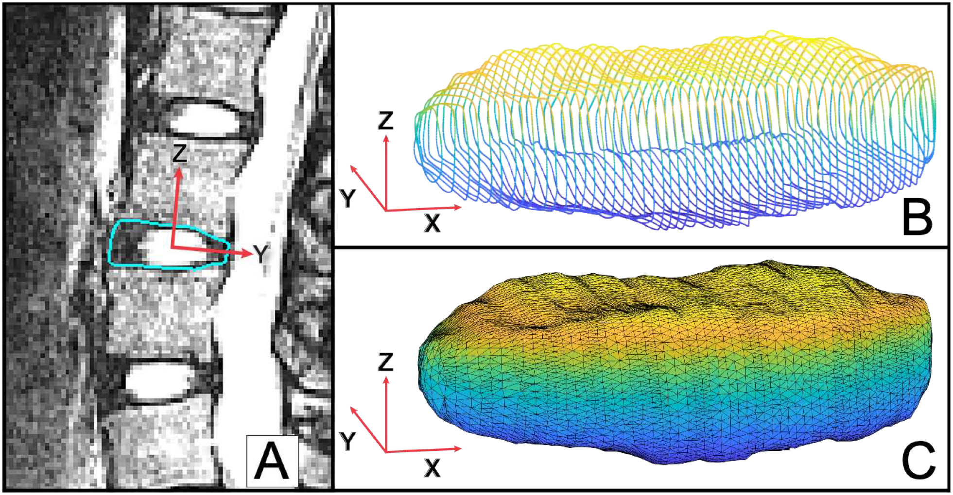
Figure 1: IVD segmentation processing.

Visualization of the directions of the 1st, 2nd and 3rd principal component vectors are denoted by the X, Y and Z axes, respectively. The Z-axis demonstrates the axis along which height is calculated. (A) The outer contours of each IVD were segmented on each slice of the MR images. (B) Segmentation yielded a 3D wireframe model of each IVD. (C) Following alignment of the 3rd principal component vector with the z-axis of the cartesian coordinate system, a 3D triangulation mesh of the disc was created for subsequent analyses.
