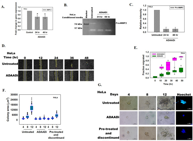
Fig 5. ADAADi treatment inhibits migration and invasion of cells.
(A). qPCR analysis of MMP-2 expression in untreated and ADAADi treated HeLa cells. (B). Zymography assay showing the secretion of Pro-MMP2 in the media by HeLa cells. (C). Quantitation of Pro-MMP2 was done using Image J software. The data was normalized with respect to untreated control and is presented as average ± s.d. of two independent experiments. (D). Image analysis of the wound assay captured at different time points after induction of gap in monolayer of HeLa cells. (E). Quantitation of the migration of HeLa cells in the absence and presence of ADAADi as a function of time. The data was normalized with respect to untreated control and is presented as average ± s.d. of three independent experiments. (F). Area (μm2) of the HeLa colonies calculated as a function of time in the absence and presence of ADAADi. (G). Colony formation monitored in untreated, ADAADi treated, and ADAADi pre-treated followed by discontinuation of the inhibitor in HeLa cells. On the 12th day, the colonies were fixed using 100% methanol and stained with Hoechst. Images were taken using Nikon TiS microscope.

