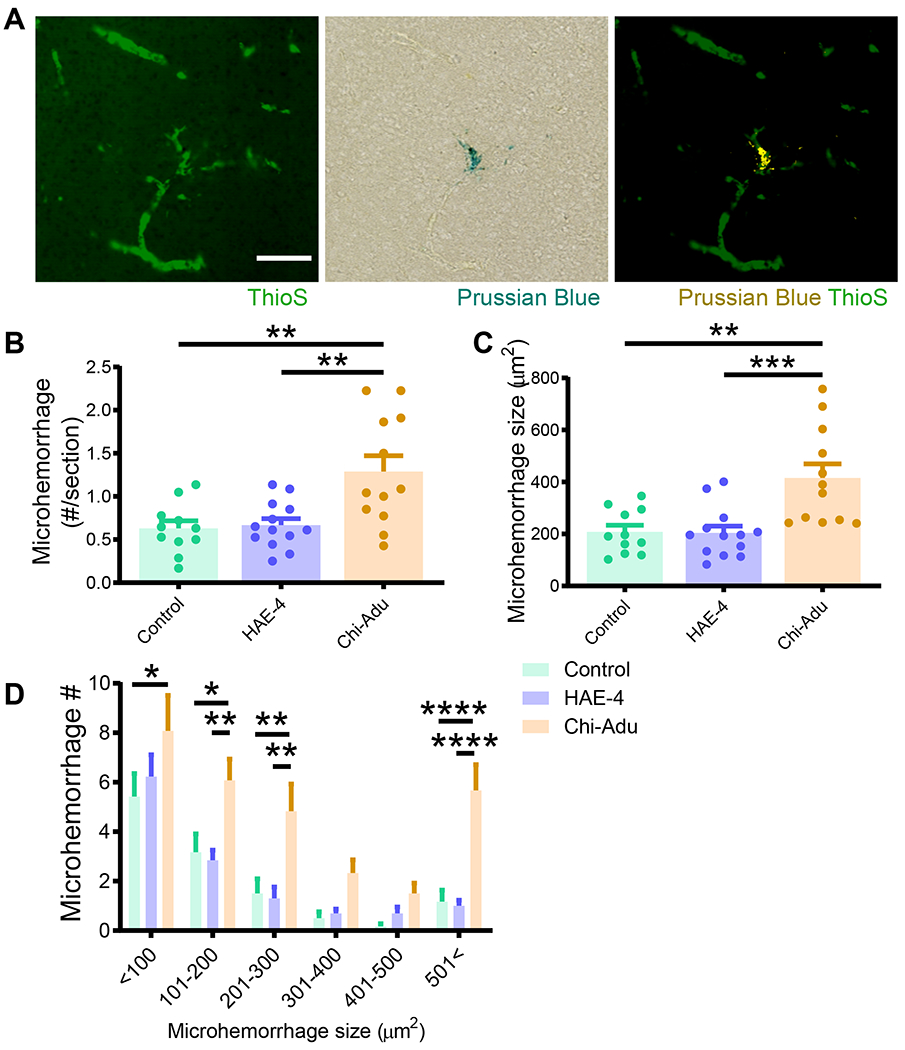
Fig. 3: Chi-Adu exacerbates CAA-associated brain microhemorrhages whereas HAE-4 does not.

A, Dual labeling of ThioS+ fibrillar CAA and Prussian blue for hemosiderin deposits revealed CAA-associated microhemorrhages (right panel: microhemorrhage digitally converted to yellow) in 10-month-old 5XE4 mice. Scale bar = 100 μm. B, C, Prussian blue staining analysis for microhemorrhage frequency (B) and size (C) in 5XE4 mice dosed weekly for 8 weeks with antibody treatment (50 mg/kg, i.p.; Control IgG, n = 11; HAE-4, n = 13, chi-Adu, n = 12). One-way ANOVA with Tukey’s post hoc test (two-sided). D, Number of microhemorrhages binned in 100 μm2 increments for frequency/size distribution. Control = Control IgG. Chi-Adu = Chimeric Aducanumab. Data expressed as mean ± SEM, two-way ANOVA with Tukey’s post hoc test (two-sided). *P < 0.05, **P < 0.01, ***P < 0.001, ****P < 0.0001. No other statistical comparisons are significant unless indicated.
