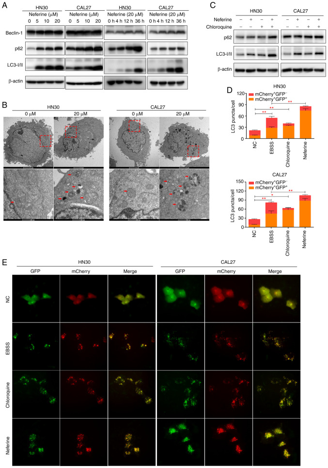
Figure 3.
Neferine promotes the generation of autophagosomes while inhibiting autophagic influx in head and neck squamous cell carcinoma cells. (A) The expression of Beclin-1, p62, and LC3 as analyzed by western blotting in HN30 and CAL27 cells following treatment with neferine at the indicated concentrations and durations of time. (B) Transmission electron microscopy of the neferine-treated (20 µM) cells depicting the number of vacuoles containing cytoplasmic materials and multivesicular bodies, a characteristic feature of degradative autophagic vacuoles. (C) Western blot analysis of p62 and LC3 expression in the HN30 and CAL27 cells concomitantly treated with neferine (20 µM) and chloroquine (10 µM). (D and E) Autophagic flux examined through double-labeled fluorescent LC3 adenovirus (mCherry+GFP−-LC3 and mCherry+GFP+-LC3). Colocalization (yellow) of both GFP (green) and mCherry (red) fluorescence indicated the generation of autophagosomes (neferine 10 µM, 24 h; EBSS 6 h; chloroquine 10 µM, 24 h). *P<0.05 and **P<0.01.

