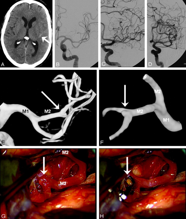
Fig 2.

A 48-year-old man presenting 4 days after headache of sudden onset in good clinical condition. A, CT scan shows a small amount of blood in the left Sylvian fissure (arrow). B–D, DSA in 3 projections fails to show a middle cerebral artery aneurysm. E and F, 3DRA depicts a 1-mm aneurysm on a M2-M3 junction (arrow). G and H, Operative view (compare with F) before (G) and after (H) clipping. Blood remnants are proof of rupture of the small aneurysm.
