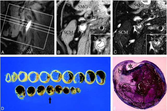
Fig 2.
A–E, An example of MR images and histopathologic features of atherosclerotic plaque consisting of LC with intraplaque hemorrhage. First, a long-axis image of the carotid artery including a stenotic lesion was obtained (A), and based on this image, 3 T1-weighted and 3 T2-weighted short-axis images of the artery were obtained for plaque characterization. B–C, Axial T1- (B) and T2- (C) weighted images corresponding to the intermediate section in the long-axis image (A). Inset, a magnification of the atherosclerotic carotid plaque. A manual operator-defined ROI was drawn on a workstation to refer to the histologic features (a, LC-IPH(+); b, FC), and the rSI at the corresponding location on the matched MR image was measured. The rSI for LC-IPH(+) on T1- and T2-weighted images were 1.89 and 0.85, respectively. The rSI for FC on T1- and T2-weighted images were 0.71 and 0.53 in this case. SCM indicates sternocleidomastoid muscle; SMG, submandibular gland; asterisk, lumen. D, Gross sections of carotid endarterectomy specimen at approximately 2-mm intervals. A section corresponding to axial MR images (B–C) is marked with an arrow. E, Masson trichrome–stained cross section matched to axial MR images (B–C) showed an abundant LC-IPH(+) and FC (a and b, manual operator-defined ROIs for LC-IPH(+) (a) and FC (b).

