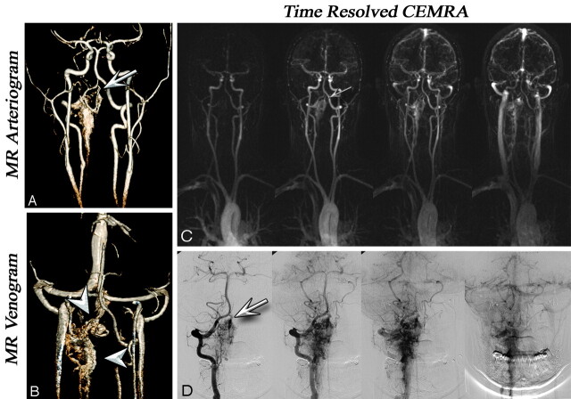
Fig 1.
A and B, Coronal VR projections from CE-MRA (voxel volume, 0.8 × 0.7 × 0.8 mm3) in a 22-year-old woman with a history of intraspinal hemorrhage and cervical laminectomy. Arterial (A) and venous (B) phase images show a diffuse serpiginous cervical C2–C3 AVM. Note the feeding vessel derived from the right vertebral artery (arrows). Venous drainage was through the paravertebral venous plexus and the occipital sinus (arrowheads). C, Time-resolved images with 1.5-second temporal resolution and 1.2 × 1 mm in-plane resolution show enhancement of the AVM via the right vertebral artery (arrows) and subsequent venous drainage. D, A phase-by-phase comparison with CCA is presented. MRA and CCA examinations were performed on the same day, before radiosurgery.

