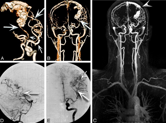
Fig 3.
A 27-year-old man status postembolization and radiosurgery for a left parieto-occipital AVM in 2003 who presented for follow-up study and assessment for repeat radiosurgery. A–E, High-spatial-resolution MRA images and corresponding CCA images are as follows: right lateral oblique (A) and coronal 3D VR images (B), coronal full-volume MIP (C), lateral projection from superselection and angiogram of the basilar artery (D), and coronal view from superselection and angiogram of the left internal carotid artery (E). Feeder arteries are identified by both techniques as angular branches of the left MCA and parieto-occipital branches of the left posterior cerebral artery (arrows). Single venous drainage into the superior sagittal sinus is identified by both techniques (arrowhead).

