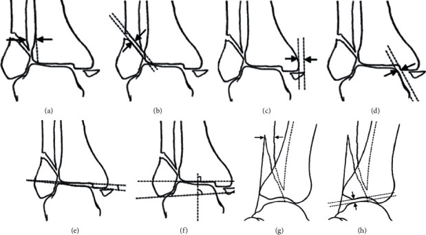
Figure 7.

Illustrations of reduction evaluation parameters. AP radiograph: (a) the width of syndesmosis; (b) the lateral displacements (LMD); (c) the displacement of the medial malleolar fragment (MMD); (d) the distance of medial clear space (MCS); (e) talar tilt angle (TTA); (f) talocrural angle (TCA). LAT radiograph: (g) the lateral displacements (LMD); (h) the anterior talar translation (ATT). This picture is partially referenced by the citations [31, 32].
