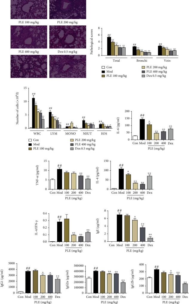
Figure 1.

PLE exerted anti-inflammation effect in allergic asthma in vivo. (a) Representative images showing pathological changes in lung tissues determined by H&E staining (magnification, ×100). (b) The scores of inflammatory cell infiltration in lung tissues (n = 15 independent slices from three mice of each group). (c) WBC, LYM, MONO, NEUT, and EOS counts in BALF (n = 8). (d–g) The level of IL-6, TNF-α, and IL-4 and the ratio of IL-4/IFN-γ in BALF (n = 8). (h–k) The concentration of IgE, IgG1, IgG2a, and IgG2b in serum (n = 8). ##P < 0.01vs. the control group; ∗P < 0.05 and ∗∗P < 0.01vs. the model group.
