
Fig. 2.
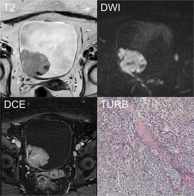
Example of correctly classified muscle-invasive BC. A 90-year-old woman with hematuria and a bladder mass reported after flexible cystoscopy underwent mp-MRI before primary TURB. T2W imaging (T2) showing an exophytic lesion on the right lateral wall, > 1 cm in the major axis with focal interruption of the SI of the muscularis propria. The b1000-DWI (DWI) confirming the high signal intensity of the tumor extending to the muscular layer. Based on bp-MRI, all readers assigned a VI-RADS category of 4. DCE imaging (DCE) showing early enhancement of the lesion and inner layer with an early enhancement of the muscularis propria, indicating tumor infiltration. DCE did not affect the VI-RADS category. T stage after TURB was HG-T2 (TURB). DCE, dynamic contrast-enhanced; DWI, diffusion-weighted imaging; HG, high grade; mp-MRI, multiparametric magnetic resonance imaging; bp-MRI, biparametric magnetic resonance imaging; SI, signal intensity; T2W, T2-weighted; TURB, trans-urethral resection of the bladder; VI-RADS, Vesical Imaging-Reporting and Data System