FIGURE 4.

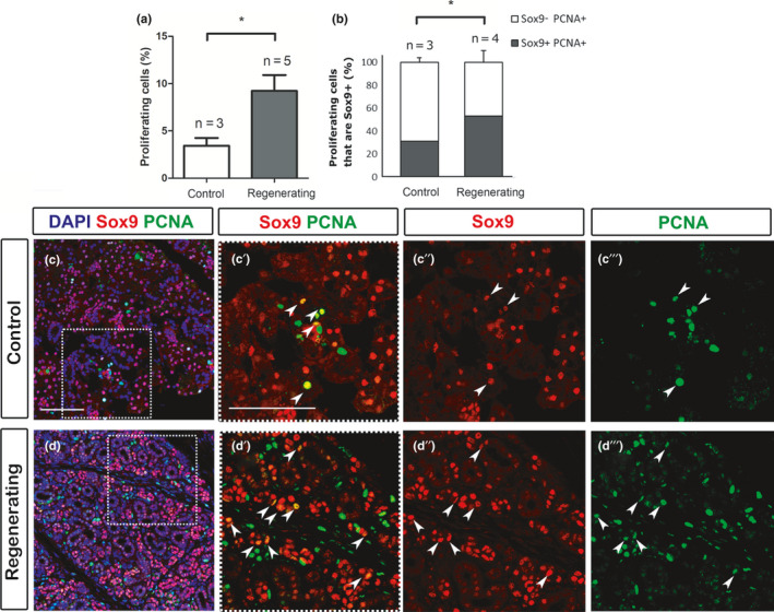
Sox9‐positive cells are highly proliferative in the regenerating submandibular gland. (a) Percentage of proliferating cells PCNA+/DAPI in the gland. (b) Percentage of proliferating cells that are Sox9‐positive (Sox9+PCNA+) and (Sox9−PCNA+) in the control unoperated and regenerating submandibular gland. Experiment follows the schedule shown in Figure 3a) with the ligation removed after 8 days and the animal culled 4 days later. N = number of mice. *p < 0.05. Error bars are s.e.m. (c, d) Immunofluorescence of Sox9 (pink) and PCNA (green). DNA is shown in blue (DAPI). All Sox9 expression is nuclear. (c–c′′′) Control unoperated. (d–d′′′) Regenerating submandibular gland. Dotted boxes in (c, d) indicate the magnified areas in c′, d′ respectively. (c′, c″, d′, d″) Sox9 (red) and (c′, c′′′, d′, d′′′) PCNA (green). Arrowheads indicate the Sox9+PCNA+ cells (yellow in c′,d′). All mice were female 6–10 weeks old. Scale bars in (c): 100 μm. Same scale in d. Scale bar in c′: 100 μm. Same scale in c″, c′′′, d′, d″, d′′′
