FIGURE 6.

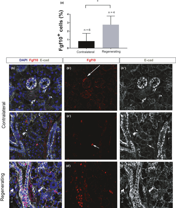
Fgf10‐positive cells increase in the ductal epithelium during regeneration. (a) Percentage of Fgf10‐positive cells in control and regenerating submandibular glands (% of total cells). *p < 0.01. (b, c) Immunofluorescence in contralateral (b, b′, b″ and c, c′, c″), and regenerating (d, d′, d″) female submandibular glands. (b, b″, c, c″, d, d″) Epithelium outlined with Ecadherin (white). DNA is shown in blue (DAPI). (b, b′, c, c′, d, d′) Fgf10 immuno (red). (b, b′, b″) Only a few Fgf10‐positive cells are identified in the duct epithelium (arrow). (c, c′ c″) Sparce Fgf10 cells overlap with E‐cadherin (arrow) indicating expression in the epithelium. (d, d′, d″) Multiple Fgf10‐positive cells are identified during regeneration, the majority located in the striated/granular ducts. All mice were female 6–10 weeks old
