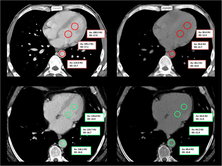
Fig. 2.
Placement of circular region of interest in the left ventricle and the descending aorta in a 42-year-old anemic male patient (serum hemoglobin 5.6 g/dl; upper row) and in a 59-year-old healthy male patient (serum hemoglobin 14.5 g/dl; lower row) in contrast-enhanced portal venous phase scans (left) and the corresponding virtual non-contrast reconstructions (right). Of note, the “interventricular septum sign” is slightly visible in the virtual non-contrast reconstruction in the upper row

