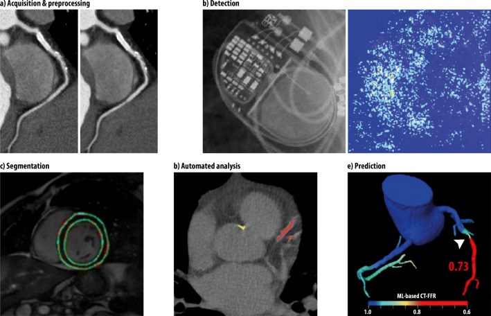
Fig. 5.
Examples of application of ML in cardiac radiology. a Curved multiplanar reformation of CTAs with multiple plaques and a stent in the right coronary artery; standard hybrid iterative image reconstruction on the left, image processed with an ML algorithm with reduced noise on the right [21]. b Correctly identified Advisa device on a plain radiograph (left) with the according saliency map (right) that visualizes the neural networks attention [22]. c Segmentation of the LV on MRI by a DCNN with automatically detected contours in green color [23]. d Automated detection and quantification of calcifications on non-contrast CT scans (red: left anterior descending coronary artery; green: left circumflex coronary artery; yellow: thoracic aorta) [24]. e ML-based CT fractional flow reserve predicting obstructive stenosis in the mid left anterior descending coronary artery [25]

