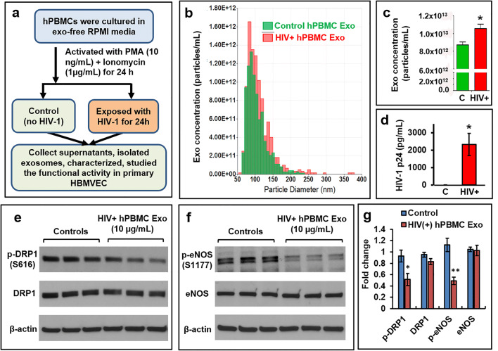
Fig. 7.
Exosomes isolated from HIV-1 infected hPBMCs reduce the expression of p-DRP1, and p-eNOS in primary HBMVECs. a The schematic was indicated the culture, activation, infection, and isolation of exosomes from either HIV-1 infected (HIV+hPBMC Exo) or control hPBMCs. b Extracellular vesicles (EVs) were isolated from HIV-1 infected and uninfected hPBMCs, and the distribution of concentrations (particles/mL) with the particle diameter (nm) was measured by qNano-IZON system. c The concentration of exosomes isolated from HIV-1 infected and uninfected hPBMCs was compared. d The presence of HIV-1 p24 protein in control Exo (C) and HIV+hPBMC Exo was measured by ELISA. Representative western blot of e p-DRP1 and total DRP1, and f p-eNOS and total eNOS in cells exposed to control Exo and HIV+hPBMC Exo. g Quantitative analysis of p-DRP1, total DRP1, p-eNOS, and total eNOS. Error bars show mean ± SD, n = 3 independent experiments, and significant changes are presented as p values (*p < 0.05, **p < 0.003)

