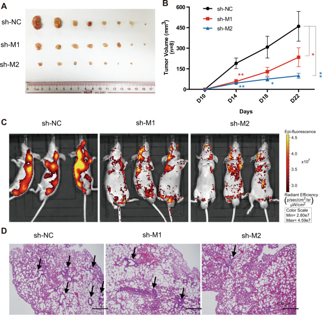
Fig. 5. MALAT1 promotes HCC tumor growth and metastasis in vivo.
A Subcutaneous tumor formation in male BALB/c-nu mice. shMALAT1-1 or shMALAT1-2 Bel7402 stable cells were injected subcutaneous on the right side of mice, while the same number of corresponding control cells were injected on the left side of the same mice. After 4 weeks, the mice were dissected, and the tumors grown under the skin were removed, measured, and photographed. B The tumor volumes were measured on day 10, 14, 18, and 22 after subcutaneous injection. n = 8/group. C In vivo metastasis assay of Bel7402 cells with MALAT1 downregulation was performed by tail intravenous injection. Representative images of mice with the scramble control (shNC), shMALAT1-1, or shMALAT1-2 knockdown constructs were taken by the in vivo animal imaging system. The bioluminescence signals indicated the location of cancer cells. D Mice after the bioluminescence signal detection were sacrificed and dissected. Organs were excised and the metastasis in lung was further investigated by immunohistochemistry analysis. Representative H&E staining images showing lung metastasis in control and shMALAT1 mice. Lesions in lung are indicated by black arrows. Scale bar represents 100 μm. Quantitative data were represented as mean ± SD. *P < 0.05, **P < 0.01.

