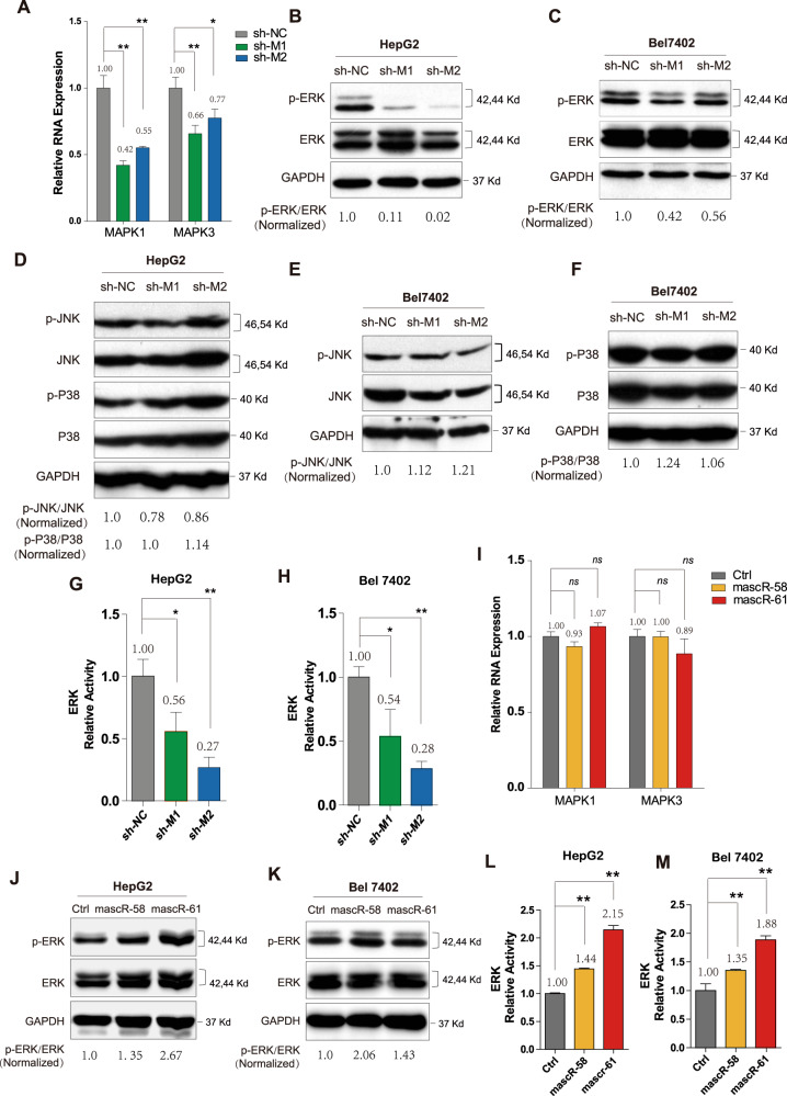
Fig. 6. mascRNA and MALAT1 active ERK/MAPK signaling pathway in HCC.
A The expressions of MAPK1 and MAPK3 were detected by real-time RT-PCR in control and shMALAT1 HepG2 stable cell lines. Western blotting for ERK and p-ERK in (B) HepG2 and (C) Bel7402 control and shMALAT1 stable cells. D Western blotting for JNK, p-JNK, P38, p-P38 in HepG2 control and shMALAT1 stable cells. Western blotting for (E) JNK, p-JNK, F P38, p-P38 in Bel7402 control and shMALAT1 stable cells. The activity of ERK signaling was examined by the dual-luciferase reporter assay in (G) HepG2 and (H) Bel7402 control and shMALAT1 stable cells. I The expressions of MAPK1 and MAPK3 were detected by real-time RT-PCR in control and mascRNA overexpressed HepG2 stable cell lines. Western blotting for ERK and p-ERK in (J) HepG2 and (K) Bel7402 control and mascRNA overexpressed stable cells. The activity of ERK signaling was examined by the dual-luciferase reporter assay in (L) HepG2 and (M) Bel7402 control and mascRNA overexpressed stable cells. All western blotting in this figure used GAPDH as a control. The band density of ERK, p-ERK, JNK, p-JNK, P38, and p-P38 were normalized to respective GAPDH, and the values of p-ERK/ERK, p-JNK/JNK, and p-P38/P38 were showed in the figure. Quantitative data were represented as mean ± SD. *P < 0.05, **P < 0.01.

