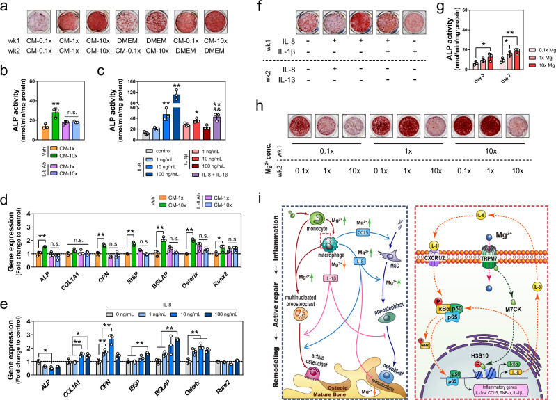
Fig. 7. The effects of Mg2+ and its modulated inflammatory microenvironment on osteogenesis.
a Alizarin Red staining of mineralized nodules of MSC treated with conditional medium from Mg2+-treated macrophages at either early or late stage of osteogenic induction. b The ALP activity of MSC cultured in conditional medium from macrophages with or without the addition of IL-8 neutralizing antibody (n = 3). c The ALP activity of MSC cultured in medium supplemented with recombinant human IL-8 or IL-1β (n = 3). d The osteogenic-related gene expression of MSC cultured in conditional medium from macrophages with or without the addition of IL-8 neutralizing antibody (n = 3). e The osteogenic-related gene expression of MSC cultured in medium supplemented with different concentrations of recombinant human IL-8 (n = 3). f Alizarin Red staining of mineralized nodules of MSC treated with recombinant human IL-8 or IL-1β. g The ALP activity of MSC cultured in DMEM supplemented with different concentrations of Mg2+ (n = 3). h Alizarin Red staining showing the mineralization of MSC treated with different concentrations of Mg2+ at either early or late stage of osteogenic induction. i The schematic shows the mechanism in which Mg2+ modulates both macrophages and mesenchymal stem cells in the bone healing process. Data are mean ± s.d. n.s. P > 0.05, *P < 0.05, **P < 0.01 by one-way ANOVA with Tukey’s post hoc test (b, c) or two-way ANOVA with Tukey’s post hoc test (d, e, g).

