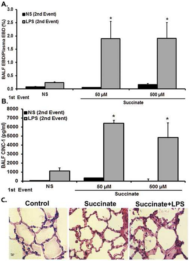
Figure 3. Succinate/LPS-induced lung injury.
Sprague-Dawley rats were transfused with 50-500 μM Succinate followed by an infusion of LPS (100 μg/kg) 6 hours later. Blood and BALF were taken to measure ALI. a) Succinate/LPS induces ALI, as measured by increased levels of EBD extravasation into BALF fluid compared to NS/NS, succinate/NS, NS/LPS (*=p<0.01 compared to all other groups). NS/LPS resulted in increased EBD compared to NS/NS and succinate/NS, but was not statistically significant (p>0.05). b) Succinate/LPS induces ALI, as measured in increased levels of BALF CINC-1 compared to NS/NS, Succinate/NS, NS/LPS (*p<0.01 compared to all other groups). No other groups show statistically significant differences. c) Representative images of lung sections stained with H&E from control (NS/NS), succinate/NS and succinate/LPS treatment groups are shown (40x). Control and succinate [50 μM]/NS animals showed little derangement in cellular architecture. The succinate [50 μM]/LPS group shows prominent neutrophil infiltration, pulmonary alveolar wall thickening, and arcuate inflammation.

