FIGURE 5.

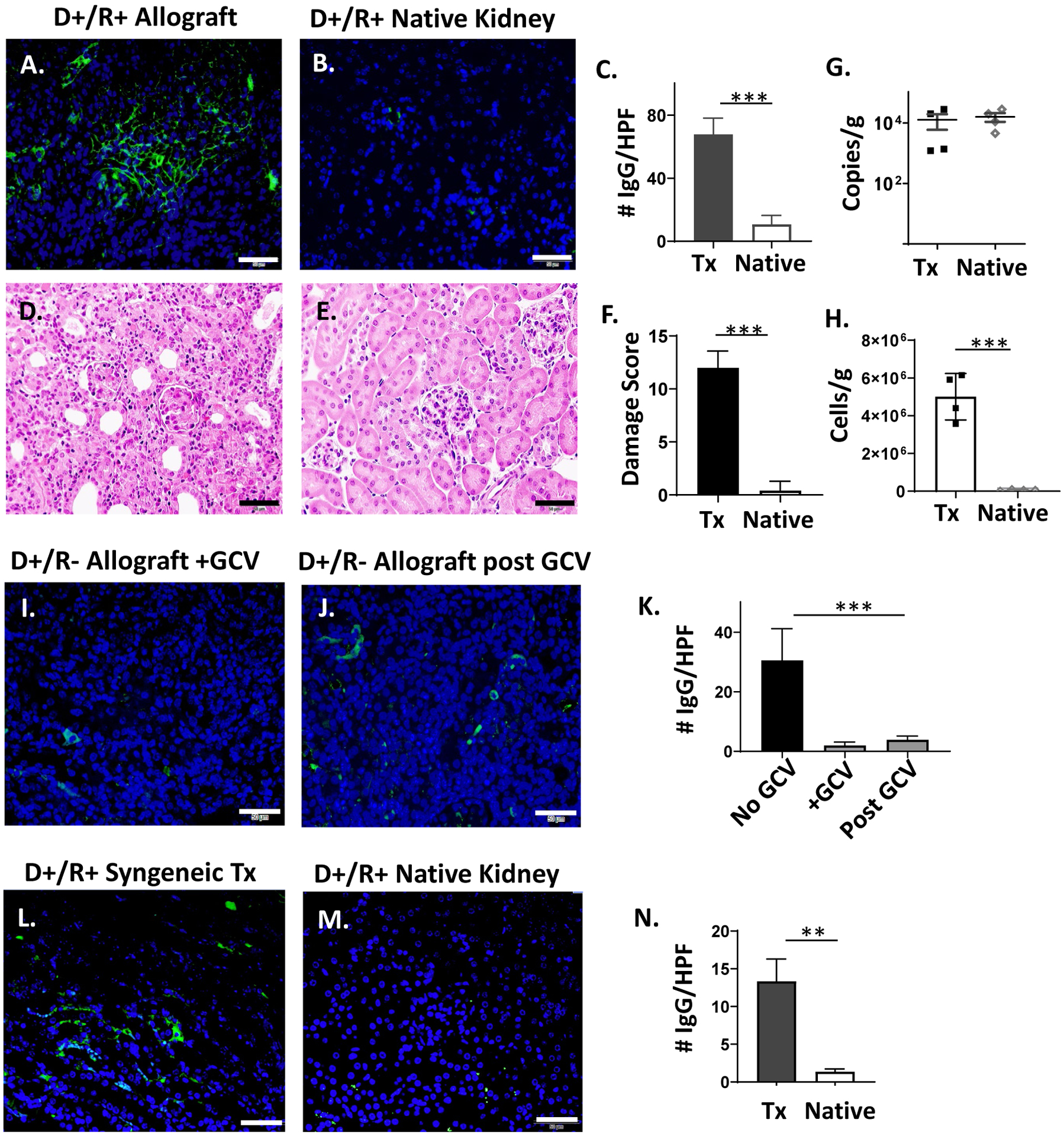
IgG immunostaining of D+/R+ native kidneys and D+/R− allografts treated with ganciclovir.
(A-B) Allografts and native kidneys of D+/R+ recipients were stained with IgG-AF488 as in Fig. 1. (C) IgG immunostaining was quantitated for 10 HPF and compared between transplant (Tx) and native kidneys (n=4/group). (D-F) Hematoxylin and eosin staining of allografts (D) and native kidneys (E) were scored for organ injury, and damage scores (F) compared between Tx and native kidneys. (G) MCMV viral loads in Tx and native kidneys were compared by quantitative DNA PCR. (H) CD45+ cell infiltrates in Tx and native kidneys were quantified by flow cytometry. (I-K) D+/R− recipients were treated with ganciclovir (GCV) for 14 days (I), or for 14 days followed by 7 days without antiviral treatment (J). Allografts were stained for IgG-AF488 (I-J) and quantitated (K) using Image J in comparison with IgG staining of day 14 D+/R− allografts without ganciclovir treatment (Fig. 3). (L-N) D+/R+ syngeneic grafts and native kidneys of immunosuppressed recipients were stained for IgG and quantitated using Image J (n=3).
(A-J) Representative images are shown (40x, bar=50 μm).
** p<0.01; *** p<0.001.
