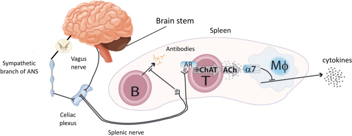
Fig. 3.
Illustration of the inflammatory reflex (here, in the spleen). Efferent signals from the brain stem travel through the efferent vagus nerve to the celiac plexus, which also receives input from the sympathetic branch. The catecholaminergic splenic nerve arises in this celiac plexus and projects inside the spleen, where its terminal fibers reach T and B lymphocytes. Efferent signals in the vagus nerve activate the splenic nerve, which releases norepinephrine in the spleen, activating the choline acetyltransferase‐expressing T-lymphocytes (ChAT) through their adrenergic receptors (AR). ChAT then release acetylcholine (ACh), which acts on α7 subunits (α7) of the nicotinic acetylcholine receptor on macrophages (MΦ), suppressing further release of tumor necrosis factors (TNF). Activation of the splenic nerve also stops B‐lymphocyte migration and inhibits their antibody production. Adapted from [111]

