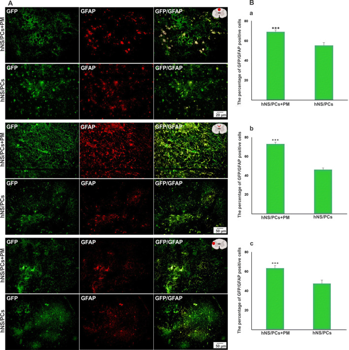
Fig. 10.
The expression of GFAP (an astrocyte marker) within the lesion site, in the perilesional region, and in the healthy host tissue (extralesional region) 8 weeks after transplantation of human neural stem/progenitor cells (hNS/PCs) with and without PuraMatrix (PM) in rats with spinal cord injury. Representative immunofluorescence images of the expression of green fluorescent protein (GFP; shown in green, left column), GFAP (shown in red, middle column), and the merged images (right column) of the hNS/PCs and hNS/PCs + PM groups in three investigated areas (a). The mean percentage of GFP-GFAP positive cells in the hNS/PCs + PM and hNS/PCs groups in three studied regions (b). Data are represented as the mean ± SD. * indicates P < 0.05

