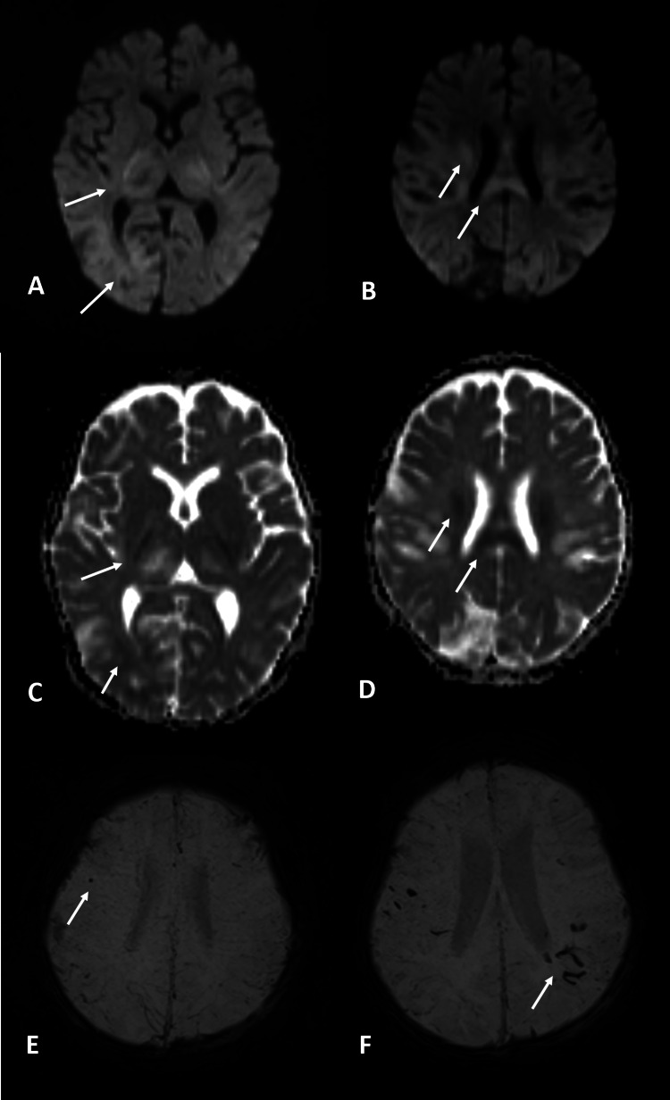
Figure 2. MRI – diffusion and SWI.
Diffusion (A, B) and ADC map (C, D) at the initial exam show restriction in the posterior limb of the internal capsule, cortico-subcortical junction of the occipital lobes (A and C), the posterior body of the corpus callosum, and corona radiate (B and D). A little dot of hemorrhage was visualized in the frontal lobe at the initial exam as a blooming artifact in the susceptibility-weighted image (E). In the follow-up exam, more hemorrhagic changes were visualized in the parietal lobes (F)
MRI: magnetic resonance imaging; SWI: susceptibility-weighted imaging; ADC: apparent diffusion coefficient

