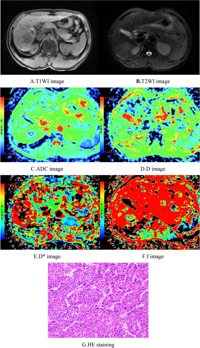
Figure 2.
Male, 61 years old, HCC in the right hepatic lobe. (A) T1WI image. The lesion showed a low signal and a relatively clear boundary. (B) T2WI image.The lesion showed a slightly high signal, with a relatively clear boundary. (C–F) ADC, D, D*, and f images, respectively. The ADC value was 1.18 × 10⁻³ mm2/s, the D value was 0.92 × 10⁻³ mm2/s, the D* value was 30.8 × 10⁻³ mm2/s, and the f value was 0.18. G. HE staining (*100) showed moderately differentiated HCC tissues.

