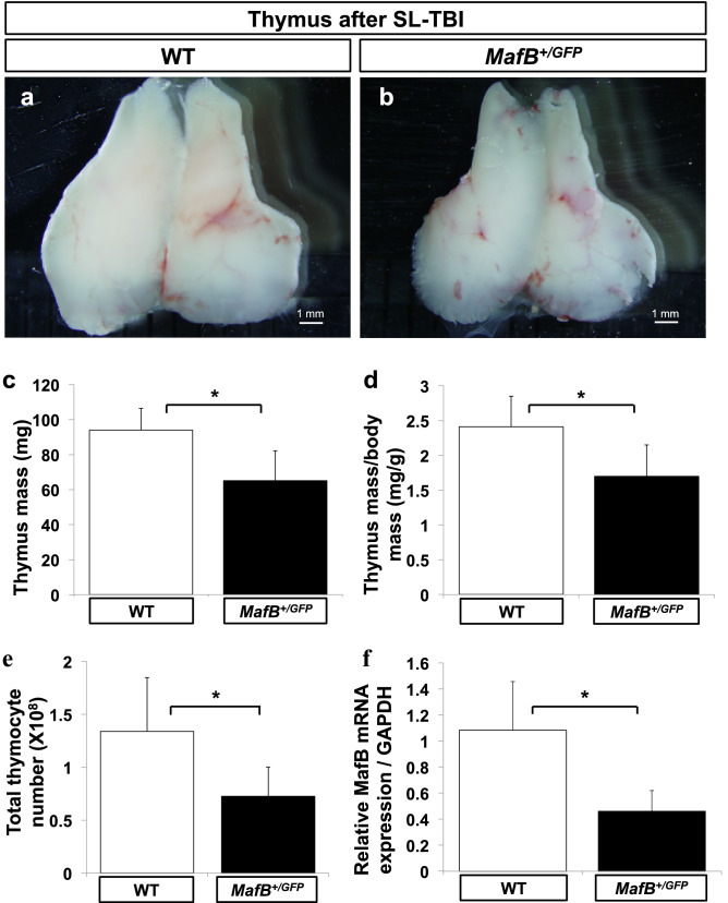
Figure 3.
MafB+/GFP mice showed impaired thymic recovery after sublethal total body irradiation (SL-TBI). (a–e) Thymi from 9-week-old wild-type (WT) (white bars, n = 6) and MafB+/GFP (black bars, n = 6) mice were analyzed 28 days after SL-TBI. (a,b) Gross morphology of thymus. Scale bar: 1 mm. (c) Absolute thymus mass (mg; milligrams). (d) Normalized mass calculated as thymic mass divided by body mass (mg/g). (e) Total thymocyte number, measured using a haemocytometer. All data are shown as the means ± standard deviation (SD). *P < 0.05 (Student’s t test). (f) The thymi of MafB+/GFP mice showed reduced MafB mRNA expression compared to those of WT counterparts. Total RNA was extracted from whole thymi of untreated 5-week-old WT (white bar, n = 6) and MafB+/GFP (black bar, n = 6) mice. MafB mRNA expression was subsequently measured by qRT-PCR, and normalized to GAPDH mRNA expression. Data presented as the means ± SD. *P < 0.05 (Student’s t test). All data shown are representative results of at least 3 independent experiments using adult specimens from different litters (n ≥ 3).

