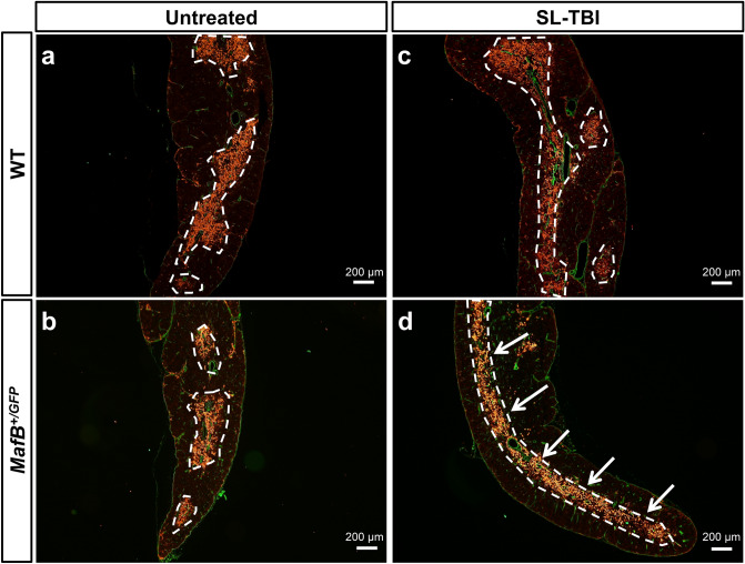
Figure 5.
Thymi from MafB+/GFP mice displayed aberrant organization of medullary thymic epithelial cell (mTEC) clusters after SL-TBI. (a–d) ER-TR7 expression (green color, representing fibroblasts and other mesenchymal cells of the thymic capsule) and Keratin 14 expression (red color, representing mTECs) shown by immunofluorescence staining of 9-week-old MafB+/GFP and WT thymi frozen sections (transverse), 28 days after SL-TBI. The borders of Keratin 14 positive mTEC clusters are indicated by white dotted lines. Scale bar: 200 µm. Similar organization of Keratin 14 positive mTEC clusters was observed in (a) untreated WT thymi, (b) untreated MafB+/GFP thymi, and (c) SL-TBI-treated WT thymi. (d) Aberrant organization of mTEC clusters was prominently observed in SL-TBI-treated MafB+/GFP thymi, which often exhibited a single large mTEC cluster (indicated by white arrows). All data shown are representative results of 3 independent experiments using adult specimens from different litters (n = 3).

