Figure 6.
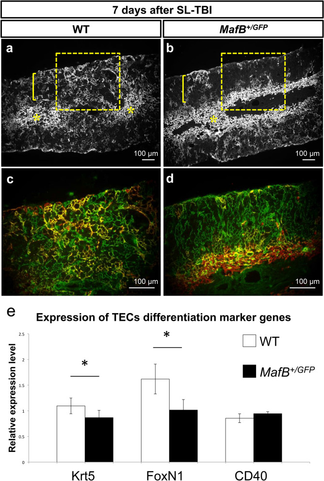
Cortical keratin 5/8 double positive cells were less prominently detected after SL-TBI; abnormal immature TECs marker expression in MafB mutant mice thymi. (a–d) Immunofluorescence staining of 6-week-old MafB+/GFP and WT thymi frozen sections (transverse), 7 days after SL-TBI. (a,b) Low magnification images showing Keratin 5 expression (white color, representing mTECs). Yellow asterisks indicate the medulla regions, whereas yellow brackets indicate cortex regions. Enclosed areas include parts of the cortex, cortico-medullary junction (CMJ) and medulla regions. Scale bar: 100 µm. (c,d) High magnification images showing both Keratin 8 expression (green color, representing cTECs) and Keratin 5 expression (red color, mTECs) of enclosed areas in (a,b). Co-localization of Keratin 5 and Keratin 8 expression is indicated by yellow color. Scale bar: 100 µm. (a,b) Keratin 5 positive cells were detected in the medulla regions of both MafB+/GFP and WT thymi. Such cells were also present in the cortex regions of WT thymi, but were less apparent in the cortex regions of MafB+/GFP thymi. (c) Cells expressing both Keratin 5 and Keratin 8 were prominently detected in the cortex of WT thymi. (d) Keratin 5/8 double positive cells were less prominently detected in the cortex of MafB+/GFP thymi. All data shown are representative results of 3 independent experiments using adult specimens from different litters (n = 3). (e) The expression of TECs differentiation marker genes after irradiation. Krt5 and FoxN1 expressions were reduced in MafB+/GFP mutant compared with those of WT. CD40 is expressed in mature TECs in controls and such expression was not altered in mutants. *P < 0.05 (Student’s t test).

