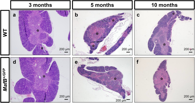
Figure 7.
MafB+/GFP mice showed signs of accelerated age-related thymic involution. (a–f) Hematoxylin and Eosin (H.E.) staining of 3, 5 and 10-month-old MafB+/GFP and WT thymi paraffin sections (transverse). Scale bar: 200 µm. (a,b) 3 and 5-month-old WT thymi showed normal thymic architecture, judged by the presence of medullary region with a clear distinction between cortex (purple-colored regions) and medulla (pink-colored regions). Black asterisks indicate medullary region. (c) 10-month-old WT thymi displayed signs of age-related thymic involution, with slightly reduced number of medullary region and slight loss of distinction between cortex and medulla (relatively small intermixed pink and purple regions). (a,d) The thymi of 3-month-old MafB+/GFP mice were histologically similar to those of WT littermates. (b,c,e,f) The thymi of 5-month-old and 10-month-old MafB+/GFP mice exhibited slightly decreased number of medullary region compared to those of WT littermates. In contrast to WT counterparts, mutant thymi also displayed a greater loss of distinction between cortex and medulla, wherein a larger portion of the tissue area is occupied by medulla regions (pink) interspersed with cortex regions (purple). These observations suggest that MafB+/GFP thymi showed signs of accelerated age-related thymic involution compared to WT thymi. All data shown are representative results of 3 independent experiments using adult specimens from different litters (n = 3).

