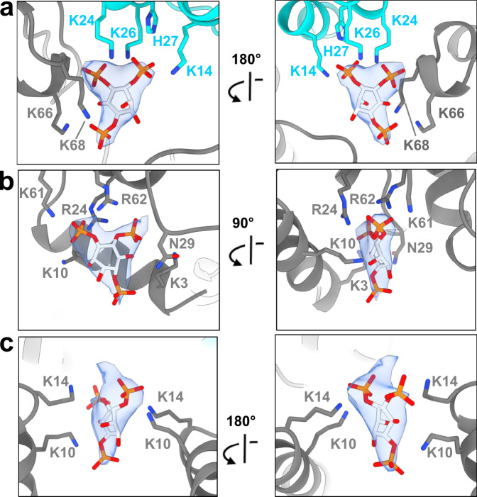
Fig. 2. PIP2 binding sites in the AENTH tetramer.
a PIP2 binding site shared by the ANTH and ENTH domains. b PIP2 binding site within the ENTH domain. c PIP2 binding site in the interface between two ENTH domains. The residues involved in PIP2 binding are shown in sticks and colored blue for nitrogen and gray and cyan for carbon in ENTH and ANTH, respectively. The polar head of the PIP2 is shown in stick format and colored red, orange, and light gray for oxygen, phosphate, and carbon, respectively with the corresponding density shown as a transparent surface.

