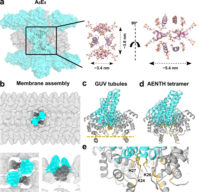
Fig. 5. Protein-lipid interactions of the AENTH 16-mer complex structure and of the ANTH-ENTH assembly on GUVs.
a, left, All the polar heads from the PIP2 placed in the structure (shown in sticks) are contained in the region near to the core of the structure (shown in surface representation), indicating the presence of a PIP2 micelle in the center of the map. Right, only the α0 helices from ENTH subunits are shown in cartoon representation (in pink) together with the PIP2 shown in stick (colored by atom), all of them are pointing towards the interior of the structure. b–e Flexible fitting of the tetrameric model to the previously obtained cryo-EM map of ANTH-ENTH coat on GUV tubules18. b Overview of the GUVs tubular coat structure with the tetramer fitting inside the lobes of the structure (EM-DB entry: EM-2896). Bottom, close-up view of the tetramer fitted into the EM density of the GUV structure. c AENTH tetramer structure fitted to the GUVs coat structure. The regions involved in PIP2 contacts are colored in gold in the cartoon representation. The bilayer plane is indicated as a gold dashed line. d AENTH tetramer in the 16-mer ANTH-ENTH complex, with the lipid-binding regions highlighted in gold in the cartoon representation. e Close-up for the ANTH lysine patch residues involved in the coordination of the polar head of PIP2 lipid.

