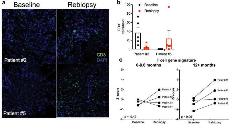
Fig. 5. T-cell content upon treatment with EGFR TKI in human lung tumor biopsies.
a Single representative images of 2 matched lung tumor biopsies submitted to immunofluorescence staining for CD3 is shown. b Five fields per biopsy were counted and the individual determinations and their mean are plotted. c An immune cell signature developed by Bindea et al.37 to deconvolute bulk gene expression data was used to infer T-cell content in the RNAseq data. Z scores for signatures predicting T cells were binned by TTP of 0–8.6 months or >12 months. Matched Z scores for each patient are graphed at baseline and upon re-biopsy and analyzed by paired t-test. The p values for the TTP of 0–8.6 months and >12 months are 0.49 and 0.06, respectively.

