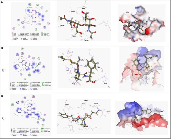
Figure 3.
(A) High-resolution crystal structures of coronavirus target explain the native ligand (NAG) in the active pocket (PDB ID: 6VW1, Score = −4.4, RMSD = 1.3). (B) High-resolution crystal structures of coronavirus target explain Alacepril in the active pocket (PDB ID: 6VW1, Score = −5.1, RMSD = 1.3). (C) High-resolution crystal structures of coronavirus target explain Lisinopril in the active pocket (PDB ID: 6VW1, Score = −4.6, RMSD = 1.3). N.B: The surface and maps representations show the H-bond donor, H-bond acceptor, and hydrophobic regions around the docked compound.

