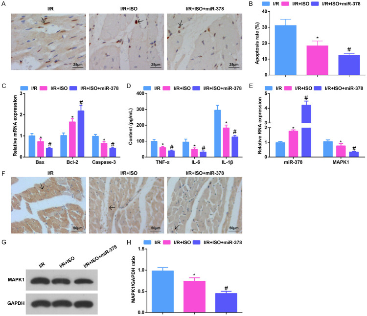
Figure 4.
Elevating miR-378 further strengthens the ISO-mediated effects on apoptosis rate and inflammatory infiltration in MI/RI mice. A. TUNEL staining results in MI/RI mice after ISO treatment and up-regulating miR-378 (× 400; Scale bar = 25 μm); B. The apoptosis of cardiomyocytes in myocardial tissues detected via TUNEL staining; C. Bax, Bcl-2 and Caspase-3 mRNA expression in myocardial tissues in MI/RI mice after ISO treatment and up-regulating miR-378 detected via RT-qPCR; D. The expression of TNF-α, IL-6, and IL-1β in serum in MI/RI mice after ISO treatment and up-regulating miR-378 detected via ELISA; E. The expression of miR-378 and MAPK1 in MI/RI mice after ISO treatment and up-regulating miR-378 detected via RT-qPCR; F. CD45 immunohistochemistry results in MI/RI mice after ISO treatment and up-regulating miR-378 (× 200; Scale bar = 50 μm); G. Protein bands of MAPK1; H. The protein expression of MAPK1 in MI/RI mice after ISO treatment and up-regulating miR-378; * vs the I/R group, P < 0.05; # vs the I/R + ISO group, P < 0.05; 5 mice in each group. The data were expressed in the form of mean ± standard deviation. One-way ANOVA was used for data analysis, and Tukey’s post hoc test was used for pairwise comparison after ANOVA analysis.

