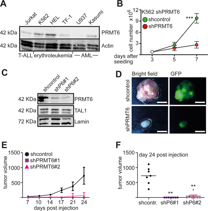
Fig. 1. PRMT6 knockdown decreases proliferation of hematopoietic cells lines.
A Western blot analysis of PRMT6 expression in Jurkat, K562, HEL, TF-1, U937, and Kasumi cells. Western blot was done with extracts from the indicated cells and specific antibodies against PRMT6. Lamin served as loading control. B PRMT6 mediates enhanced proliferation. PRMT6 was knocked down by shRNA in K562 cells. Six days after transduction shPRMT6 and shcontrol cells were seeded out in similar numbers. Cells were counted at the indicated time points. The error bars display the standard deviation from the mean from three determinations. The P-values were calculated using ANOVA. ***P < 0.001. C Western blot showing efficient knockdown of PRMT6 with shRNA. Western blot against the transcription factor TAL1 and against actin served as controls. These cells were injected subcutaneously into C57BL/6 mice. D Analysis of subcutaneous tumors upon injection of shcontrol and shPRMT6 K562 cells, respectively. Bright field and GFP image of an exemplary tumor from day 24 is displayed. The white bar indicates 0.5 cm. E Tumor growth curve from day 7 after injection until day 24 is shown for two shPRMT6 constructs. Tumor volume is given in mm3. F Endpoint analysis at day 24 of post-injection. Tumor volume is given. The P-values were calculated using ANOVA from seven mice. **P < 0.01, ***P < 0.001.

