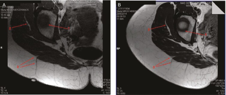
Figure 7.
Magnetic resonance imaging of cellulite from this 29-year-old woman at (A) baseline and (B) after subcision.60 Baseline image (A) shows a clear spot on the top of the depressed lesion with a perpendicular thick fibrous septum associated with this lesion and (B) the same area 7 months after subcision, showing the severed septum. Arrows 1 and 2 indicate anatomic structures utilized as a guide to obtain the same slices of bone and muscle layer, respectively. Arrow 3 points to the septum arising from the muscle. Reprinted with permission from Hexsel et al.60

